


Technologies
ABOUT MCLINIC
TECHNOLOGIES
Our modern practice has eight treatment rooms and two operating rooms, which are optimally adapted to the requirements of surgical procedures. In addition, we have high-quality technical equipment, including a three-dimensional X-ray (DVT /CBCT), three-dimensional stereo photogrammetry (VECTRA™) and virtual surgery planning options (3D Surgery Dolphin Imaging™).
We also ensure the highest standard of hygiene with detailed documentation in our clinic-level sterilization room and throughout our practice.
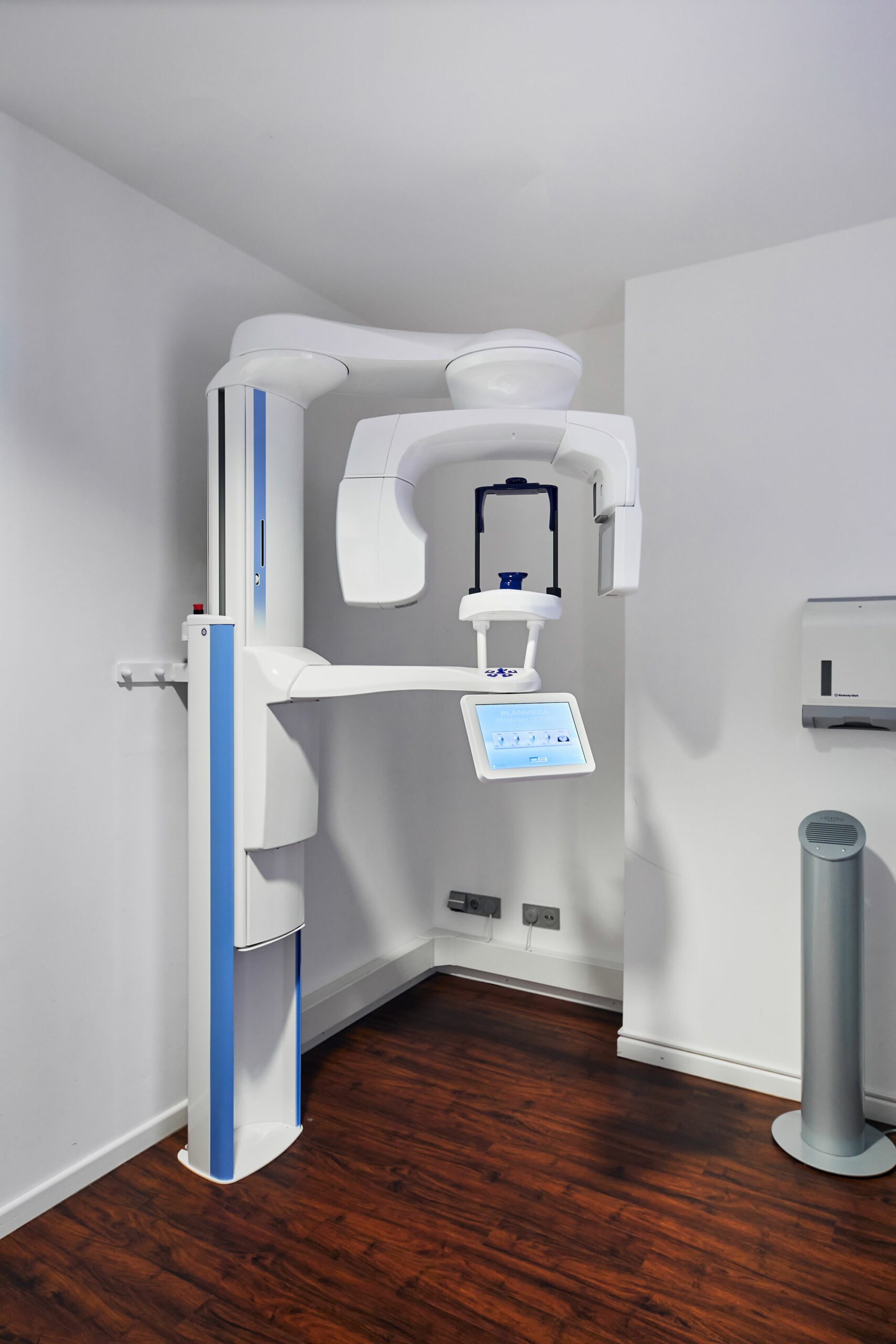
The digital volume tomography unit (DVT), also known as the cone-beam computer tomography (CBCT) is a high-tech X-ray device that was developed specifically for the facial region. It can be used to create three-dimensional images, especially of the jaws and facial skeleton. The main advantage of DVT images is that they enable much more precise and meaningful diagnostics than conventional X-ray images.
For example, it is often only possible to see the exact width and condition of the jawbone or to assess the spatial course of an important nerve pathway by means of 3D imaging. This allows us to get a more accurate picture of the anatomical conditions, since the anatomical structures are also shown in depth. Furthermore, the images are particularly detailed, true to scale and undistorted.
In general, treatments such as implantations, wisdom tooth removals and jaw relocations can be planned more precisely and thus usually performed more safely and gently. Lastly, compared to the alternative computed tomography (CT), DVT significantly reduces radiation.
At MFACE, we work with the Planmeca ProMax 3D Mid, an all-in-one DVT unit that offers 3D imaging, 3D photos, 2D digital panoramic and cephalometric radiography.
How DVT works:
- DVT provides individual cross-sectional images of the jaw or entire face.
- These individual images can either be used directly for true-to-scale measurement or combined into a complete image with the help of a computer program.
- The complete image is then available on the monitor for computer-assisted 3D surgery planning (implantation, wisdom tooth removal, jaw relocation).
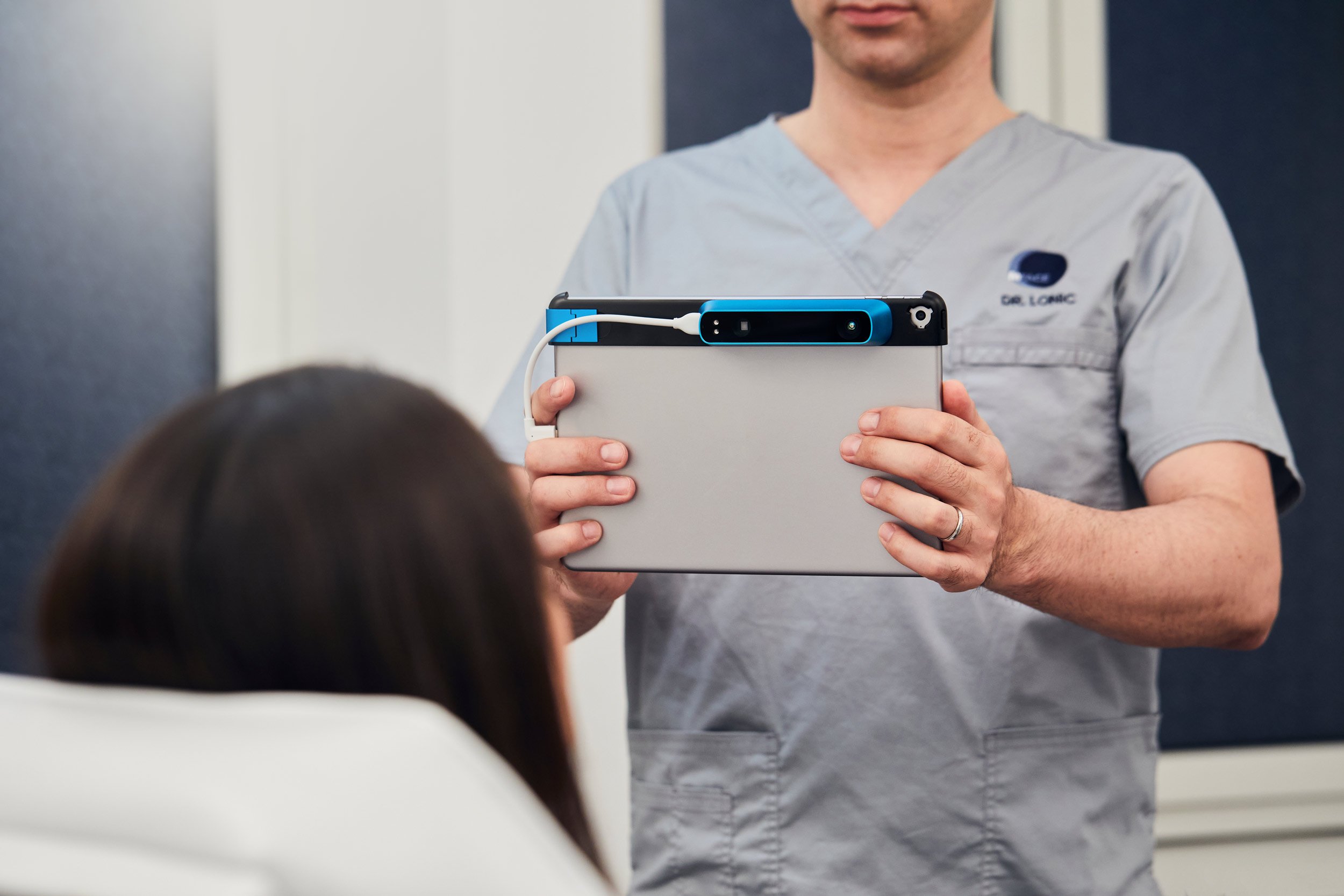
The VECTRA™ Stereophotogrammetry is the latest generation of imaging technology at our disposal. The face, breast and body surface is captured by a high-end camera by taking three photographs. From the data set, a 3D image is generated in a matter of seconds, which serves as the basis for planning aesthetic, functional and reconstructive procedures in the respective areas.
Data sets from DVT or CT can be overlaid with the VECTRA™ system, resulting in a precise representation of bony and soft tissues that elevates surgical planning, especially in the area of dysgnathic surgery, to the highest possible level. Also, aesthetic procedures such as rhinoplasty and facial fat injections can be extensively visualized and possible outcomes can be illustrated to our patients in real time.In breast surgery, different implant sizes can be simulated to ensure the right size for Your desired outcome. This is done without any radiation exposure.
The planning of surgical measures in the facial region is millimeter work. Conventional 2D planning cannot offer this precision due to superimposition of the bony structures, especially in more complex cases; therefore, we plan our orthognathic procedures (dysgnathia) based on the three-dimensional DVT and VECTRA data using so-called “virtual surgery” with Dolphin 3D Surgery™.
Here, the movements of the bony segments and the soft tissues can be simulated on the computer, thus increasing safety for the patient and the surgeon.
Modern 3D diagnostics do not only allow an exact diagnosis, but also enable a virtual surgery simulation. This computer-assisted navigated surgery offers patients the greatest accuracy and safety.
The virtual surgery plan is precisely translated into navigation aids such as drilling templates, which combined with the surgeon’s experience guarantees an optimal surgical result.
Water-jet assisted liposuction (WAL) is a gentle body contouring procedure. A special ultra-thin cannula containing a rinsing fluid is inserted into the fatty tissue with pulsating bursts of spray and immediately aspirated along with the fat cells. Due to the simultaneous infiltration and suction of the fan-like water jet, a time-consuming infiltration of the area is no longer necessary.
Results are quickly visible due to the gentle procedure, as swelling is significantly less with this procedure. The difference between this method and conventional liposuction is that the fat cells are suctioned off with much less force and thus with a considerably reduced risk of side effects. In addition, this method requires less pain medication.
The operating time is significantly reduced. Also, the fat can be collected at the same time and used for injections to other parts of the body (face, breast, buttocks).
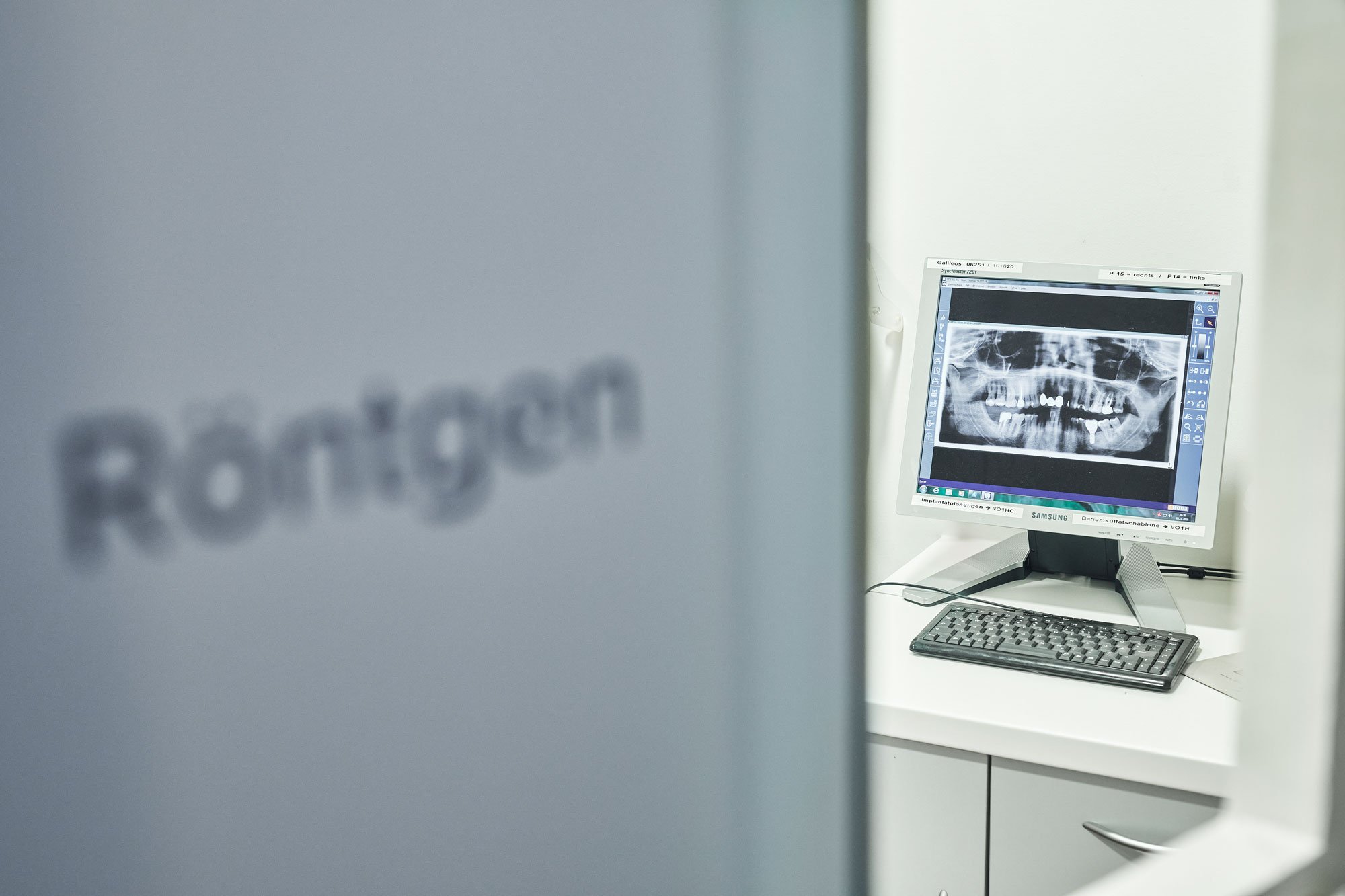
In our practice we use the most modern type of X-ray examination – digital X-ray. Until now, conventional X-ray was associated with higher radiation exposure for the patient. Digital X-rays enable us to perform all your X-rays with up to 90% less radiation exposure.
Your safety and health are our top priority. Our modern X-ray and computer system enables us to immediately display and process the X-ray images taken on the monitor. The high resolution and the possibility to process the digitized images afterwards guarantees an accurate and safe diagnosis and gives us and you the possibility to work out an exact treatment plan together based on the display on the monitor.
The advanced technology makes photo development unnecessary. This protects the environment and saves you waiting time. In addition, digital files are always quickly available and can be transferred by e-mail to any point on earth in seconds.
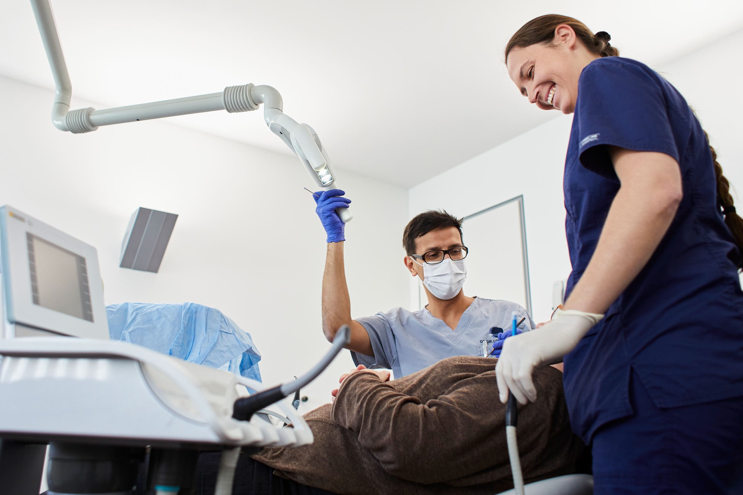
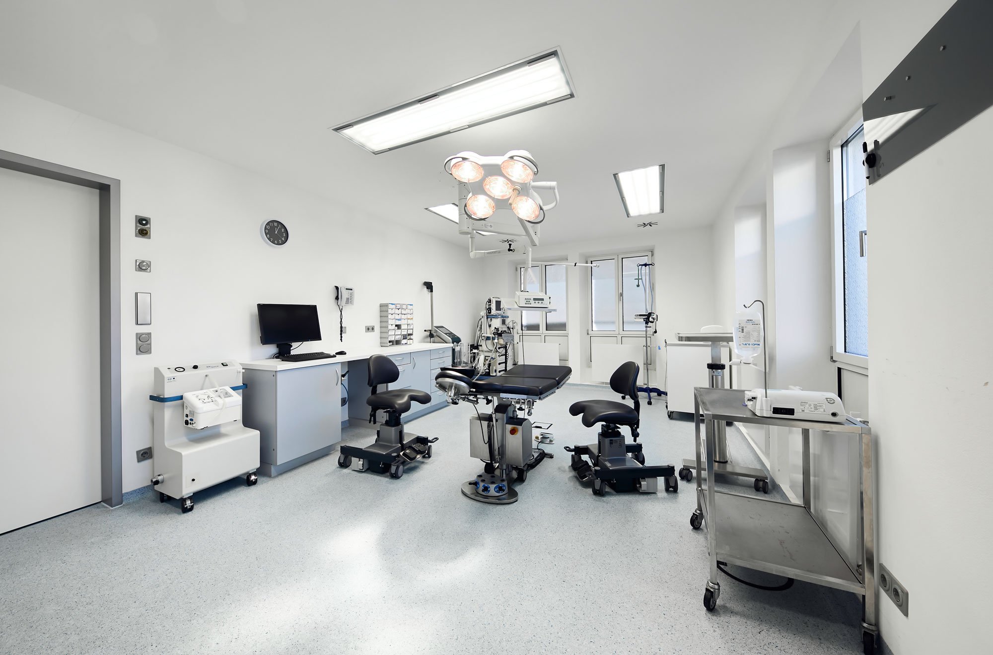
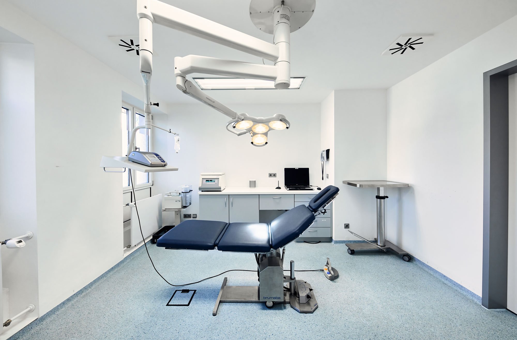
Surgical treatment success depends not only on the experience and care of the surgeon, but is also determined by optimal general conditions such as hygiene and sterility. For the safety of our patients, we therefore perform all operations not in treatment rooms or procedure rooms but in our integrated clinic in two highly modern class I operating rooms.
This means that we comply with the recommendations of the German Society for Oral and Maxillofacial Surgery and the Robert Koch Institute for Hygiene. These operating rooms are characterized by high requirements for low germ levels in the operating room. This is achieved through elaborate structural measures such as room air systems and airlocks, which are otherwise only provided by hospitals.
Through these measures, we guarantee a maximum of low germ count and thus ensure your safety and the success of your operation.
We would rather invest the time that used to be spent on cumbersome administrative tasks in the care of our patients. That is why our practice and clinic are networked and all data and findings are stored digitally on a central server. All treatment rooms of the practice, the operating rooms of the clinic, the reception, the X-ray, the sterilization, the technology and the offices are connected in a powerful network.
X-ray images and findings, as well as surgery documentation and sterilization data, can be retrieved at any time from any workstation. The documentation of the course of treatment and the constant availability of all data supports our planning and ensures the safety of your treatments.
Piezosurgery technology is used for implantation or bone augmentation and represents a revolution in bone surgery. The use of this device as an “intelligent” scalpel ensures high precision in bone preparation and safety for nerves and vessels.
The technology is based on ultrasound; the waves are modulated and thus their intensity is precisely matched to the types of tissue to be worked on.
“How do I look after a breast augmentation, after a filler injection, or after a nose job?” This question is on the minds of many patients. VECTRA is the world’s leading 3D imaging solution for cosmetic surgery, which can be used to virtually simulate such procedures and model the body parts to be changed according to the patient’s ideas. From three simple photographs in our consultation, the powerful software can generate a 3D image reconstructing the corresponding body region within seconds.
Using the simulation tools, the doctor can perform very specific modeling on the virtual body area. In the field of breast surgery, in addition to determining the shape, volume and position of the breast implant, it is possible, for example, to raise the breast, reduce the size of the areola or even – especially if you wish to have a breast lift – simulate the anticipated scars. On the basis of the simulation we can better visualize your wishes (e.g. size of the implants, positioning, shape of the tip of the nose, etc.) and in this way obtain a surgery plan which illustrates the possible result and can aid your decision for the operation. In addition, many problems can be identified before the operation and included in the surgical planning at an early stage.
We are
here for you
MAKE AN APPOINTMENT ONLINE
Just a few clicks to your individual desired appointment. Simply enter your type of insurance, your reason for treatment and your preferred appointment time.
Opening HOURS
Our opening hours are Monday to Thursday from 8:00 AM – 5:00 PM, and on Fridays from 8:00 AM – 2:00 PM, as well as by appointment.
CONTACT
Our online reception, including our digital telephone assistant, is available for you 24/7 at +49 (0)89 8292 440 or at any time with the adresses of the respective departments.
HOW TO FIND US
You can reach us by car and by local and long-distance public transport. Our premises are conveniently located near the Munich-Pasing train station.
HOW TO FIND US
You can reach us by car and by local and long-distance public transport. Our premises are conveniently located near the Munich-Pasing train station.


